


P60 Artificial Intelligence Colour Doppler Ultrasound
- Product Code: P60
- Availability: 1
SonoScape P60 - a reliable, high-performance system that uses an Artificial Intelligence Based Ultrasound Platform wiz+
The 21 “LED monitor on the articulated arm and the height-adjustable, laterally rotatable control panel enable comfortable work under a wide variety of examination conditions.
The introduction of Artificial Intelligence WIS on the P60 brings unprecedented improvements in efficiency and accuracy. With well-trained image algorithms of artificial intelligence, the structure recognition and manual measuring procedures are replaced by one-button operation.WIS has a wide range of functions with which tissue structures and lesion properties can be automatically recognized and analyzed. WIS makes the acquisition and interpretation of ultrasound images efficient, convenient and, above all, precise.
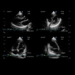
The Digital Color Doppler Ultrasound System is a general-purpose ultrasonic imaging instrument intended for use by aqualified physician for evaluation of Fetal, Abdominal, Pediatric, Small Organ (breast, testes, thyroid), Cephalic (neonataland adult), Trans-rectal, Trans-vaginal, Peripheral Vascular, Cerebral Vascular, Musculo-skeletal (Conventional andSuperficial), Cardiac (pediatric and adult), Trans-esoph.(Cardiac), Laparoscopic, OB/Gyn and Urology.
This model it is also Designed for advanced OBGYN as P60 can support advanced 4D probe
A large number of different probe types , including single crystal probes offer an optimal imaging result in all areas of application.
Device hardware
- 21.5 ″ high-resolution LED color monitor
- 13.3 ″ high-resolution touchscreen
- Height-adjustable and rotatable control panel
- Five active probe ports
- Pin probe connector
- Built-in ECG module (including hardware and software)
- WiFi module
- 1 TB hard drive
- Gel Warmer
- built-in rechargeable battery (operating time up to 2 hours without power supply)
Standard Software
- B (2B & 4B) Mode
- M Mode
- Anatomic M Mode
- Color M Mode
- Color Doppler Flow Imaging
- Power Doppler Imaging / Directional Power Doppler Imaging
- Tissues Doppler Imaging
- Pulse Wave Doppler Imaging
- Continuous Wave Doppler Imaging
- High Pulse Repeat Frequency
- Tissue Harmonic Imaging
- Pure Inversion Harmonic Imaging
- Compound Imaging
- Tissue Specific Imaging
- Image Rotation
- μ-Scan: Speckle Reduction Technology
- SR Flow
- Simultaneous Mode (Triplex)
- FreeHand 3D Imaging
- B Mode Panoramic Imaging / Color Panoramic Imaging
- Lateral Gain Compensation
- Trapezoid Imaging
- Widescan Imaging
- Biopsy Guide
- C-xlasto (Strain Elastography)
- Needle Visualization Enhancement (VIS-Needle)
- Bladder Volume Measurement
- Zoom (Pan-Zoom / HD-Zoom / Scr-Zoom)
- TEI Index
- PW Auto Trace
- Auto IMT
- Auto EF
- Auto OB: BPD / HC / AC / FL / NT / HL
- S-Guide
- Build-in User Mannual (Help)
- Sono-help (Scanning Tutorial)
- DICOM 3.0: Store / C-Store / Worklist / MPPS / Print / SR / Q&R
Optional Software :
- Micro F
- Static 3D / 4D (Must be Configured with Volumetric Probe)
- AVC Follicle (Auto Volume Calculation)
- Auto Face (Must be Configured with 3D / 4D at the Same Time)
- S-Live (S-Live / S-Live Silhouette)
- S-Depth
- Color 3D
- STIC (Spatio-Temporal Image Correlation)
- Color STIC (Spatio-Temporal Image Correlation)
- Free Vue
- VCI (Volume Contrast Imaging)
- ABD Contrast Imaging(Including TIC Analysis, MFI and MFI-Time)
- SMP Contrast Imaging
- Stress Echo
- MCE (Myocardial Contrast Echocardiography)
- LVO (Left Ventricular Opacification)
- Tissue Tracking with Quantitative Analysis (Strain Rate)
- S-Fetus: S-Fetus(acq.) & S-Fetus(meas.)
- S-Thyroid
- S-Breast
- S-MSK
- S-Follicle 2D
- S-Endo
- S-Face
- S-Spine
- Sono-Mate
- Sono-Synch
- Sono-Drop
Compatible Probes :
Linear
- Linear Probe 12L-A
- Linear Probe 12L-B
- Linear Probe 9L-A ( Low Frequency )
Convex
Endocavity ( Transvaginal )
Volume
Features
Fetal Pericallosal Artery With Micro F
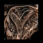
Uterus Septus With S-Live Silhouette
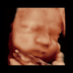
Fetal Face With S-Live
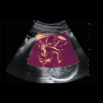
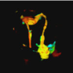
Umbilical Cord Blood Flow With Color 3D
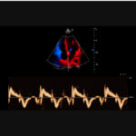
Stress Echo
Fallopian Tube With 4D HyCoSy With SPI
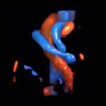
Tissue Doppler Imaging
HCC With MFI-Time
Artificial Intelligence
Brings unprecedented improvement in effectiveness and accuracy.
The adoption of AI in the P60 not only greatly simplifies the workflow, but also provides greater reproducibility and reliability in the evaluation.
S-Fetus
S-Fetus is an easy-to-use tool that enables fully automatic and accurate detection of the most relevant planes and most commonly used fetal biometry measurements.
S-thyroid
S-Thyroid is an advanced tool for diagnosing and classifying suspicious thyroid lesions, based on the ACR TI-RADS (American College of Radiology Thyroid Imaging Reporting and Data System) guideline. After selecting the region of interest, S-Thyroid can automatically define the lesion boundaries and generate a report on the characteristics of the suspected lesion.
S-Breast
S-Breast helps to delineate the borders of the lesion (simply by adjusting the ROI to the suspicious lesion) and classifies the breast lesion under study, according to the standard BI-RADS (Breast Imaging – Reporting and Data System) reports. Streamlining the workflow can improve efficiency and provide standardized reporting on the classification of benign and malignant masses.
S-MSK
S-MSK aims to solve the problems derived from the difficulty of recognizing anatomical structures in MSK, with a simple "click", the anatomical structures are highlighted and annotated on the image. Add to all this the quality of our SonoScape high frequency linear probe, and MSK diagnostics will be very easy, very hard to match.
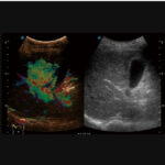
CEUS
Unlocks the full potential of microflow imaging
The P60's comprehensive contrast quantification and ultrasound imaging package offers clinicians a complete solution for assessing perfusion dynamics in a wide range of clinical settings.
Micro F
Allows visualization of microvascularized structures
Micro F provides an innovative method to extend the range of visible flow on ultrasound, especially for visualizing small, slow-flow vessels.
Advanced Cardiovascular Software
Comprehensive Solution for Cardiac Assessment
Equipped with SonoScape's unique all-glass transducers and cutting-edge technology, the P60 is committed to restoring every detail and element for accurate diagnosis.