P20 Colour Doppler Ultrasound System for Obstetrics and Gynecology
- Product Code: P20 for Obstetrics and Gynecology
- Availability: 1
P20 Colour Doppler Ultrasound System for Obstetrics and Gynecology
Slim Color doppler mid-end trolley system with 21.5" monitor with 2 transducers , compatible to HD probes and latest generation probes including single crystal probes, flexible monitor arm. Height-adjustable and turnable panel.
Incorporating innovative technologies, P20’s user-friendly designed with a simple operation panel, intuitive user interface and a variety of intelligent auxiliary scanning tools, will significantly improve your daily examination experience. Besides general imaging applications, P20 has entitled with diagnostic 4D technology which has an extraordinary performance in obstetrics and gynecology applications.
Device Hardware
- 21.5 "high-resolution LED color monitor on the multifunctional articulated arm
- Height-adjustable, rotatable, user-defined programmable control panel
- Additional 13.3 "touchscreen
- Backlit, pull-out keyboard
- Five probe slots (four active + one "parking lot")
- "Single Crystal" peeling heads with C-field beam former
- very large cine memory
- A pin probe connector
- Wireless wifi connection
- internal storage on HDD
Software:
- extensive measurement software for all departments
- 4 D image display
- 2D speckle reduction technology
- B (2B & 4B) mode
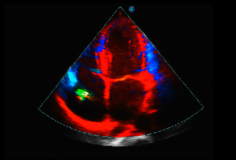
- Color Doppler flow imaging
- Pulse wave doppler imaging
- HPRF
- Continuous Wave Doppler Imaging
- Power Doppler Imaging / Directional Power Doppler Imaging
- U-scan for Speckle Reduction
- Color Doppler (Dynamic Color Technology) Triplex, PW and CW
- Trapezoid representation
- Preset management
- Auto NT
- Color Doppler panorama
- M mode
- Tissue harmonic imaging
- Tissue-specific imaging
- Pulse Inversion Harmonic Imaging
- Spatial Imaging
- LGC (Lateral Gain Compensation)
- Full screen zoom
- Image rotation
- Real-time 2D panorama recordings - 4 D image display
- ECG module
- THI Tissue Harmonic Image
- TDI (tissue doubler)
- Auto - IMT measurement
- Auto M-tuning, optimal image and Doppler settings at the push of a button
- AVC Folicle (Auto Volume Calculation )
Probes:
Linear:
- L741
- L742
- L752
Convex:
- 3C-A single Crystal Probe bdomen, obstetrics, gynecology and urology (center frequency 3.2 MHz, FOV 70°).
- C1-6
- C613
-
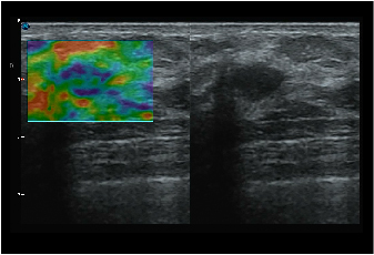
C-Xlasto Imaging:
With C-xlasto Imaging, P20 enables comprehensive quantitative elastic analysis. Meanwhile, C-xlasto on P20 is supported by linear, convex and transvaginal probes, to ensure good reproducibility and highly consistent quantitative elastic results.
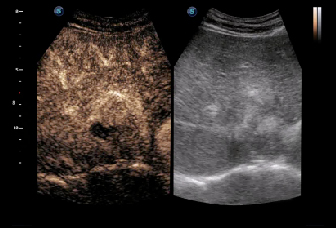
Contrast Imaging:
Contrast Imaging with 8 TIC curves allows doctors to assess perfusion dynamics in a wide range of clinical settings, including both the location and evaluation of lesion parts.
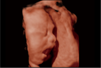
S-Live:
S-Live allows for detailed visualization of subtle anatomical features, thereby enabling intuitive diagnosis with real-time 3D images and enriching patient communication.
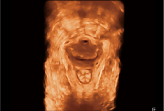
Pelvic Floor 4D:
Transperineal 4D pelvic floor ultrasound can provide useful clinical values in assessing the vaginal delivery impact on the female anterior compartment, judging whether the pelvic organs are prolapsed or not and the extent, determining if the pelvic muscles were torn accurately.
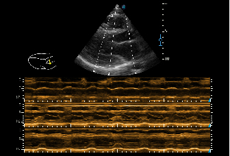
Anatomic M Mode:
Anatomic M Mode helps you observe the myocardial motion at different phases by freely placing sample lines. It accurately measures the myocardial thickness and the heart size of even difficult patients and supports the myocardial function and LV wall-motion assessment.
Tissue Doppler Imaging:
P20 is endowed with Tissue Doppler Imaging which provides velocities and other clinical information on myocardial functions, facilitating clinical doctors with the ability to analyze and compare the motions of different parts of the patient's heart.